

Types of Vision Tests: What Do They Measure?
Home / Vision Education Center /
Last Updated:
Table of Contents
Whether you have healthy or poor vision, it is important to see an eye doctor regularly for proper testing. The overall health of your vision will help your doctor determine when further intervention is necessary.
There are certain types of tests that are performed for everyone during a comprehensive eye examination, such as visual acuity testing. For some people with specific vision issues, additional testing may be performed, such as testing for color blindness.
The doctor will begin by evaluating the state of your vision and eye health. From here, they can determine which tests are appropriate for your comprehensive eye examination.
The following types of tests highlight the wide variety of vision tests that are available.
Visual Acuity Testing
Visual acuity testing is done to determine how sharp your vision is. The doctor will usually have you look at an eye chart for this examination. They will ask you to read the numbers or letters that are present on a specific line.
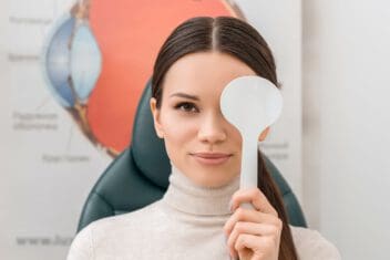
In some cases, doctors give you a handheld chart. This one is used to determine how sharp your vision is close up.
You deserve clear vision. We can help.
With 135+ locations and over 2.5 million procedures performed, our board-certified eye surgeons deliver results you can trust.
Your journey to better vision starts here.
Color Blindness Testing
The Ishihara color test is most often used to diagnose red-green color blindness. It consists of a series of colored spot pictures. A figure in the form of one or several Arabic digits is embedded in the pictures as multiple spots in slightly different colors. Individuals with normal color vision can detect the numbers, but those with color deficiencies cannot.
Another color blindness test is the Farnsworth-Munsell 100 hue test. Here you are required to arrange color chips or caps into a gradual transition of color between anchor caps. The HRR test is a red-green color test. Unlike the common Ishihara test, this test can help detect Tritan defects.
Ocular Motility Testing
This test is done to see if your eyes are moving as they should. It tests your ability to accurately focus on two separate objects and follow a moving object.
This test is important because if you have abnormal eye movements, you are at a higher risk for affected reading ability and eye strain.
Cover Test
This test allows doctors to determine your eye alignment. You will cover one eye and look across the room to see an object. This is also done on an object that is close to you.
During the test, the doctor is looking to see if you are showing symptoms of certain conditions, such as amblyopia or strabismus.
Retinoscopy
If you need a prescription for corrective lenses, a retinoscopy may be performed. The doctor is looking at the light reflection from your eye.
You will go into a dimmed room with the doctor. You will be asked to look at a specific letter on the eye chart. As you continue to stare at this letter, the doctor flips lenses and shines a light in front of your eyes. The purpose of this test is to get a lens power estimate for corrective lens prescriptions.
This test is ideal for people who are not able to sit up properly. It may also be helpful for babies and others who cannot raise their heads.
Depth Perception Test
This test is performed to evaluate your depth perception. It can also be used to determine if you are able to fully visualize three-dimensional objects.
For this test, you wear 3D glasses. There are test patterns in a booklet that you will examine. The doctor will ask you which of the circles appears closer than the others. If you choose the wrong circles, you may have a problem with your depth perception.
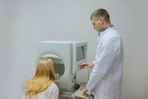
Autorefractors and Aberrometers
These are tools that doctors can use to estimate your corrective lenses prescription automatically. They are two different devices that have you look at a detailed image or a pinpoint of light.
Refraction Testing
When you go to the eye doctor to get a corrective lens prescription, this test is done to ensure your prescription is exact. The doctor uses a phoropter instrument for this test. They put it in front of your eyes while showing you a variety of lens choices.
You tell the doctor which of the lenses is the clearest. This test can be done to determine the level of farsightedness, presbyopia, nearsightedness, and astigmatism you have. Due to the level of accuracy, a doctor may prefer these tools over retinoscopy whenever possible.
You deserve clear vision. We can help.
With 135+ locations and over 2.5 million procedures performed, our board-certified eye surgeons deliver results you can trust.
Your journey to better vision starts here.
Slit Lamp Examination
This examination involves the use of a binocular microscope. The doctor uses this microscope so they can get a better visual of your internal eye structures. It offers very high magnification, making it easier to spot abnormalities that are present.
The doctor may do this test as part of a routine eye examination. They might also recommend it if they suspect certain eye diseases, such as macular degeneration, diabetic retinopathy, cataracts, and corneal ulcers.
Pupil Dilation
This is another test that doctors might use to get a better visual of your internal eye structures. The doctor will apply eye drops to your eyes that dilate your pupils. You need to wait 20 to 30 minutes after the eye drops before the doctor can do the examination.
Visual Field Testing
If a doctor suspects that you may have blind spots in your vision, this is a test they may recommend. It allows the doctor to evaluate your peripheral vision.
Blind spots or reduced peripheral vision can occur as a result of several eye or brain conditions, such as stroke complications or glaucoma.
Applanation Tonometry
Applanation tonometry is a diagnostic test that measures intraocular pressure, the fluid pressure inside your eyes. It is commonly used to assess the risk of developing glaucoma.
Eye doctors may order one to rule out or confirm glaucoma if other eye tests indicate a possible issue. Other symptoms that may necessitate applanation tonometry include blurred vision, loss of peripheral vision, tunnel vision, eye reddening and severe eye pain.
The gold standard instrument for measuring intraocular pressure is the Goldman Applanation Tonometer, which is an instrument that applies the Imbert-Fick law. This test involves gently pressing a flat-tipped probe against your eye surface. There are other applanation tonometry methods, although they are considered less accurate.
When eye pressure goes untreated, it can lead to damage to the optic nerve, which may result in vision loss. According to the American Academy of Ophthalmology, glaucoma is among the leading causes of blindness in people above the age of 60. The effects of glaucoma are usually painless and may progress for many years without your knowledge. It is for this reason that applanation tonometry is a vital eye test for detecting any changes early on.
Fluorescein Angiography
Fluorescein angiography refers to an eye exam involving a camera and special dye (fluorescent dye) to observe blood flow in the retina and choroid (two layers at the back of the eye). This test is a dye tracing technique. Hyper fluorescence or hypo fluorescence of the dye indicates certain eye problems.
Hyper fluorescence can indicate abnormal vasculature, leaking defects, eye staining, or pooling defects. Hypo fluorescence may indicate blocking defects or filling defects. This test can detect and diagnose vein occlusions, diabetic retinopathy, tumors, macular degeneration and retinal artery occlusions.
Maintaining Eye Health
Throughout your life, your vision will experience changes. Opticians and ophthalmologists recommend regular checkups even if you don’t experience any problems with your vision.
Many people require more than just vision screening. A comprehensive dilated eye exam is necessary. Certain conditions may develop for years without showing any notable symptoms. A detailed eye exam is vital for early detection and thus easier early treatment.
You deserve clear vision. We can help.
With 135+ locations and over 2.5 million procedures performed, our board-certified eye surgeons deliver results you can trust.
Your journey to better vision starts here.
References
- Color Blind Tests. All About Vision.
- Going Old School: A Refresher on Retinoscopy. Review of Optometry.
- Refraction Test. Healthline.
- How Is Eye Pressure Measured? (May 14, 2018.) Bright Focus Foundation.
- What is Glaucoma? (September 22, 2021). American Academy of Ophthalmology.
- How to measure intraocular pressure: applanation tonometry. (December 2007). Community Eye Health Journal.
- What is Fluorescein Angiography? (June 28, 2021). American Academy of Ophthalmology.
- New Farnsworth-Munsell 100 hue test norms of normal observers for each year of age 5-22 and for age decades 30-70. (December 2002). British Journal of Ophthalmology.
- The new Richmond HRR pseudoisochromatic test for colour vision is better than the ishihara test. (May 25, 2005). Clinical and Experimental Optometry.
This content is for informational purposes only. It may have been reviewed by a licensed physician, but is not intended to serve as a substitute for professional medical advice. Always consult your healthcare provider with any health concerns. For more, read our Privacy Policy and Editorial Policy.