Keratoconus: Signs, Timelines & Treatments
Home /
Last Updated:
Table of Contents
Typically, your cornea is round and dome-shaped. In some people, it can thin out and become more cone-shaped, which can be caused by the progressive disorder keratoconus. Blurry vision, sensitivity to light, swelling of the eye, and distorted vision can all be symptoms of keratoconus.
Keratoconus typically progresses slowly over several years. In the early stages, keratoconus can be treated with soft contact lenses.
As it progresses, different forms of contacts can become necessary. Procedures like corneal cross-linking can be optimal for slowing the progression of keratoconus and improving vision and corneal stability.
What Is Keratoconus?

The cornea is the front part of your eye. A normal cornea is dome-shaped and round.
Keratoconus involves the thinning and irregular shaping of your cornea, which can cause it to become more cone-like. This ends up causing vision distortion, as light is no longer focused correctly onto the retina.
You deserve clear vision. We can help.
With 135+ locations and over 2.5 million procedures performed, our board-certified eye surgeons deliver results you can trust.
Your journey to better vision starts here.
Keratoconus is likely to have a genetic component. The American Academy of Ophthalmology (AAO) reports that 10 percent of people struggling with the disorder have a parent who has it too. Eye allergies and excessive rubbing of the eyes can also be risk factors for keratoconus.
Keratoconus typically begins in adolescence or early adulthood and progresses slowly over time, usually over a period of 10 to 20 years, the American Optometric Association (AOA) explains. In some cases, it can progress rapidly and lead to corneal swelling and scarring. Keratoconus can make it hard to see clearly and ultimately requires vision correction.
Signs of Keratoconus
Early on, signs of keratoconus include:
- Blurry vision
- Sensitivity to light
- Distorted vision, causing lines to look “wavy”
- Swelling of the eye
- Eye redness
As keratoconus progresses, you may notice:
- Frequent changes in your eyeglass or contact lens prescription
- Cloudy vision
- Difficulties with night vision and driving at night due to glares
- Trouble fitting contacts in your eyes
You deserve clear vision. We can help.
With 135+ locations and over 2.5 million procedures performed, our board-certified eye surgeons deliver results you can trust.
Your journey to better vision starts here.
Keratoconus Timeline
Keratoconus can begin anywhere from age 10 to age 25, Mayo Clinic publishes. It usually progresses slowly for a period of 10 years or so.
While keratoconus starts in late adolescence to early adulthood, it continues to slowly progress up until about age 40 or so. At this time, the progression of the disorder usually stops.
Keratoconus can start out with mild vision distortion, but it can then progress into significant difficulty seeing clearly without correction. Typically, the disorder impacts both eyes, but keratoconus can progress differently in each eye.
Treatment Options
Options for treating keratoconus are going to depend on its severity and how quickly the disorder is progressing. In the early stages, eyeglasses or contact lenses can be used to correct vision and the astigmatism of the cornea. As keratoconus progresses, additional measures often need to be explored.
Corneal cross-linking is a safe and effective treatment option for keratoconus that has been shown to halt the progression of the disorder. It can also serve to improve vision. For advanced keratoconus, surgical measures may be necessary. Options include corneal implants.
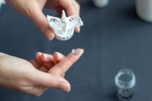
- Soft contact lenses and/or eyeglasses: Early on, eyeglasses or soft contact lenses can help to correct vision distortion and the astigmatism caused by keratoconus. Soft contact lenses can be custom-made specifically to treat keratoconus, and they are measured exactly to your eyes and specifications. Typically, contact lenses designed to treat keratoconus are larger in diameter than traditional contact lenses, to give them more stability in the eye.
- Gas permeable contact lenses: Rigid gas permeable (RGP) contact lenses can “vault” over the cornea. They may then provide a better fit than soft contact lenses for keratoconus. Unlike eyeglasses or soft contact lenses, RGP contact lenses can change the shape of your eye, smoothing it out more, to improve your vision.It can be tricky to fit contact lenses for keratoconus, however. As keratoconus progresses, your prescription can keep changing. It can even get to a point where it is uncomfortable to wear them in your eyes at all.
- Additional forms of contact lenses: There are other types of contact lenses that can be useful for treating keratoconus, such as “piggybacking” two types of contact lenses, hybrid contact lenses, or scleral lenses.
- Piggybacking contacts: This involves using both a soft contact lens and a gas permeable lens — one on top of the other. The soft lens is placed in the eye first for comfort and then the rigid lens is put on top of it to correct shape. Your eye care provider will need to make sure that enough oxygen can reach your eyes when using this method.
- Hybrid contact lenses: These contact lenses are a combination of soft and gas permeable lenses. They are rigid in the center to provide the vision and astigmatism correction of a gas permeable lens, with a comfortable outer ring that is a soft lens. Hybrid contact lenses made specifically to treat keratoconus are often more comfortable than RGP lenses, offer better vision correction than soft lenses, and are less of a hassle than piggybacking.
- Scleral contact lenses: The white part of your eye is called the Scleral contact lenses are larger than typical contacts, covering a bigger diameter of the eye and resting on the sclera. They are still gas permeable lenses, but their size keeps them from moving around as much as traditional gas permeable lenses. They can vault over the cornea better without putting as much pressure on its irregular shape.
- Corneal cross-linking: This is a procedure that was officially FDA-approved in 2016, but the journal Review of Optometry reports that it has been the chosen treatment for controlling the progression of keratoconus since its inception in 2003. Corneal cross-linking is a minimally invasive, safe, and effective outpatient treatment for keratoconus that can slow the progression of the disorder and improve vision.
Corneal cross-linking involves using liquid B2 (riboflavin) and controlled ultraviolet (UV) light to create links in the collagen fibers in your cornea. This both stabilizes and strengthens it.
This procedure can actually stop your cornea from thinning and becoming more irregular. Other treatment methods just improve vision through corrective lenses while keratoconus continues to progress. Studies show that corneal cross-linking can improve vision, stop the progression of keratoconus, and flatten the irregular shape of the cornea. Results remain stable for at least a year after the procedure.
Corneal cross-linking can also be combined with corrective eye surgery and corneal implants. A corneal cross-linking procedure can reduce the need for corneal transplants as well.
- Corneal implants: Implants, such as the thin prescription inserts Intacs, can be inserted into the mid-cornea to flatten its irregular shape to correct for keratoconus. This is a surgical procedure that has been FDA-approved to treat keratoconus.
- The procedure is quick, taking 10 minutes or so. The implants can be exchanged or removed as needed through additional procedures.
- Corneal implants, such as Intacs, can improve vision and be more comfortable than other contact lenses. However, they will not slow or stop the progression of the disorder like corneal cross-linking can.
- Corneal transplant: Advanced stages of keratoconus may require a corneal transplant. This is a surgical procedure. It is also called a penetrating keratoplasty or PKP.For this procedure, you will need a donor with a healthy cornea. Most donor corneas are from deceased individuals.
Your irregular tissue will be removed and replaced. There is a risk that your body can reject the transplant, Mayo Clinic. Even with a corneal transplant, you will often still need to wear prescription eyeglasses or contact lenses to correct your vision. You will not be able to wear contacts until your corneas are completely healed, which can take up to several weeks.
A corneal transplant is typically considered the last course of action in treating keratoconus. All other options have usually been explored and exhausted first.
You deserve clear vision. We can help.
With 135+ locations and over 2.5 million procedures performed, our board-certified eye surgeons deliver results you can trust.
Your journey to better vision starts here.
References
- What Causes Keratoconus? (December 2019). American Academy of Ophthalmology (AAO).
- Keratoconus. (2019). American Optometric Association (AOA).
- Keratoconus. (March 2019). Mayo Clinic.
- Your Top 12 Crosslinking Questions- Answered! (January 2018). Review of Optometry.
- Analysis of 2-Year Corneal Cross-Linking Results in Keratoconus Patients. (May 2014). Journal of the Egyptian Ophthalmological Association.
- Keratoconus: Causes, Symptoms and 10 Treatment Options. (July 2019). All About Vision.
- Intacs Surgery for Keratoconus. (2019). National Keratoconus Foundation (NKCF).
- Cornea Transplant. (May 2018). Mayo Clinic.
This content is for informational purposes only. It may have been reviewed by a licensed physician, but is not intended to serve as a substitute for professional medical advice. Always consult your healthcare provider with any health concerns. For more, read our Privacy Policy and Editorial Policy.