

Understanding Hyphema: Symptoms, Treatment & More
Home / Eye Conditions & Eye Diseases /
Last Updated:
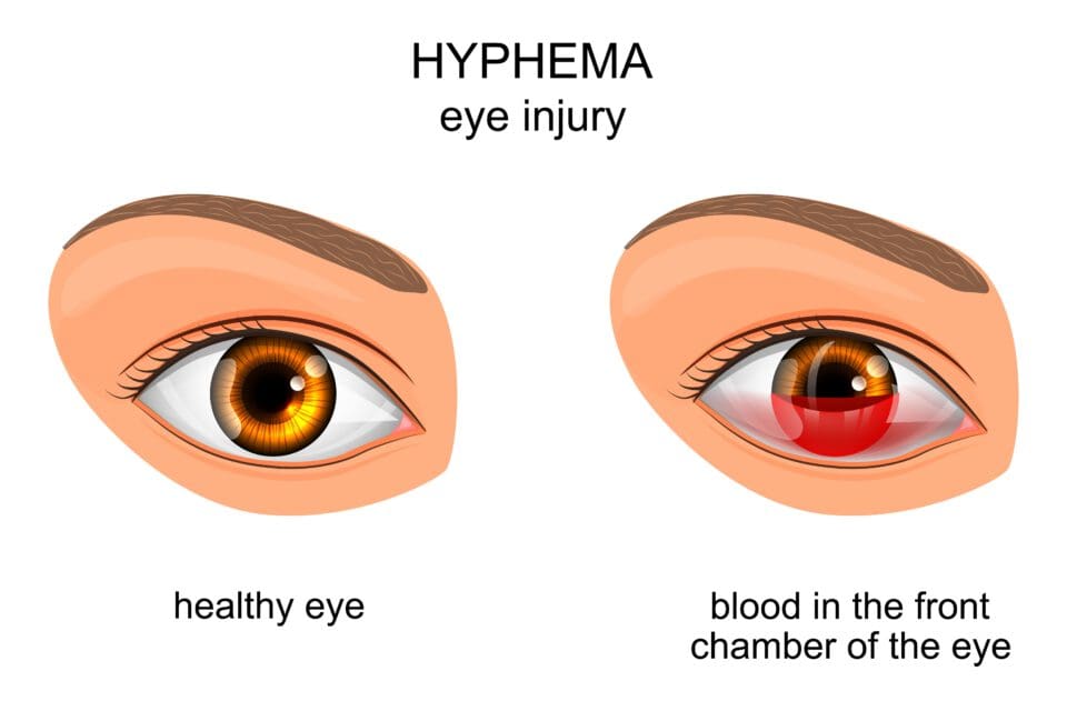
A hyphema is an eye injury characterized by a collection of blood in the eye and one that can cause discomfort, blockage of vision and other vision issues, particularly light sensitivity.
Table of Contents
Trauma is the usual cause of this condition, but there are cases where it can arise without trauma because of certain medical conditions. In most cases, permanent damage does not occur, but it is possible.
Because of the potential for long-term repercussions, people should seek prompt treatment when they see blood their eye(s). Treatment can range from prescription medications to home remedies to more extensive treatment for complications that arise. Treatment also depends on several factors, such as the person’s age, how they tolerate medications, and the severity of their injury.
There is also the risk for serious complications if a hyphema is not treated.
Doctors can perform several tests to make an accurate diagnosis. If trauma is the cause, it is also important for a doctor to screen for other potential issues, such as a concussion.
Once someone has an accurate diagnosis of a hyphema, there are treatment options.
What Is a Hyphema?
Hyphema is the presence of grossly visible blood in the anterior chamber of the eye. When you get a direct blow to your eyeball, disruption of the ocular vessels leads to the accumulation of blood between the iris and the cornea. Iris is the colored portion of your eye, while the cornea is the colorless layer covering the front part of the eye. Normally, a clear fluid known as aqueous humor fills the space between the two, giving optimal vision.
Bleeding into the anterior chamber because of trauma or an underlying medical condition causes partial or total obscuring of the pupils that interferes with vision. Even a low-grade hyphema could be associated with severe intraocular injury.
Another explanation for blood in the eye is a broken blood vessel in the eye. However, this is a separate condition known as a subconjunctival hemorrhage. This hemorrhage is usually harmless and not painful. A hyphema usually causes pain.
Without prompt and proper treatment, a hyphema can lead to permanent vision issues.
An injury to the eye that results in the pupil or iris tearing is usually responsible for this condition. Other less common factors that may cause a hyphema include:
- Abnormal surface blood vessels on the iris
- Blood clotting disorders
- Cancers of the eye (only in rare cases)
- Herpes virus-related eye infections
- Artificial lens problems following cataract surgery
Symptoms of Hyphema
Signs and symptoms that indicate you might have hyphema include:
- Bloody eye: A large hyphema will discolor the whole eye as blood replaces aqueous humor in the anterior chamber. Simple inspection is enough to identify sizeable hyphema. However, some blood pools are too tiny for the naked eye to pick.
- Visual disturbance: Vision changes will vary depending on the degree of hyphema. With pupillary obstruction, you might only be able to identify hand movements in front of the face or detect some amount of light.
- Light sensitivity: Medically referred to as photophobia, hyphema can deliver a significant sensitivity to light in which your eye begins to hurt or you experience a headache when exposed to too much light.
- Eye pain: Blood in the anterior chamber causes irritation and pressure effect that leads to eye pain.
You deserve clear vision. We can help.
With 135+ locations and over 2.5 million procedures performed, our board-certified eye surgeons deliver results you can trust.
Your journey to better vision starts here.
The severity of these symptoms depends on the degree of hyphema. Usually graded from 0-4, the symptoms worsen based on the portion of the anterior chamber filled with the blood. The grades:
- Grade 0: A microscope is necessary to see the red blood cells, but the blood pooling is not visible.
- Grade 1: The pooled blood fills less than a third of the chamber.
- Grade 2 The pooled blood fills up to half of the chamber.
- Grade 3: The pooled blood fills more than half of the chamber, but not all of it.
- Grade 4: The chamber fills completely with blood. It is called an 8-ball hyphema if the blood is dark red. It is called total hyphema if the blood is bright red.

Potential Complications of a Hyphema
Complications of hyphema are primarily attributed to the retention of blood in the anterior portion of the eye. Without appropriate intervention, you can have:
- Glaucoma: As blood pools in the eye, it leads to increased intraocular pressure, which is characteristic of glaucoma. If not treated, glaucoma can result in permanent vision loss.
- Corneal bloodstaining: Prolonged blood retention in the anterior chamber predisposes a subject to corneal staining with subsequent cloudy vision.
- Secondary hemorrhage: Severe traumatic injury often presents with recurrent bleeding that can lead to ocular tissue scarring.
- Loss of vision: Non-glaucomatous optic atrophy resulting from transient or chronic intraocular pressure elevation will cause vision loss if hyphema is not treated immediately.
Getting a Diagnosis
A patient’s complete medical history is needed to make a clear diagnosis. Doctors will want to determine if the you recently experienced any issues that could cause bleeding in the eye or trauma that affected the eye.
Your doctor will examine the eye to look for a hyphema or any signs of trauma and perform one or more tests, such as:
- Tonometry: This test is done to determine the level of pressure in the eye. There are three methods the doctor can choose from; however, the one that flattens the corneal area is the most accurate. The eyes are numbed before the procedure to alleviate discomfort. This test involves using a slit lamp, and dye is often used. There is a machine the doctor adjusts to obtain a pressure reading.
- Comprehensive eye examination: This is performed to assess a person’s ability to see.
- Eye pressure examination: Since a potential complication of a hyphema is an increase in eye pressure, this test is performed to see if the pressure is at an abnormal level.
- CT scan: If trauma was the cause of the hyphema, the doctor may order a CT scan to determine if the eye socket is fractured.
- Slit lamp test: This is done to get a better look inside the eye. It allows the doctor to use a combination of bright light and magnification so they can better assess the eye’s internal structures.
You deserve clear vision. We can help.
With 135+ locations and over 2.5 million procedures performed, our board-certified eye surgeons deliver results you can trust.
Your journey to better vision starts here.
Treatment Options
All the treatment options aim to promote blood clearing, reduce eye pressure and prevent recurrent bleeding. The majority of the cases are mild and don’t require medical intervention. That means even though you should be safe and visit your doctor, odds are the condition will abate on its own or with minimal treatment.
Any degree of hyphema could be a pointer to underlying severe intraocular injury. Therefore, you need to seek professional intervention immediately. Among the treatment options:
- Protect the affected eye by wearing a special shield over it.
- Help the eye drain by raising the head of the bed.
- Rest and avoid physical activity for the specified amount of time.
- See an ophthalmologist regularly, so they can assess eye pressure and healing.
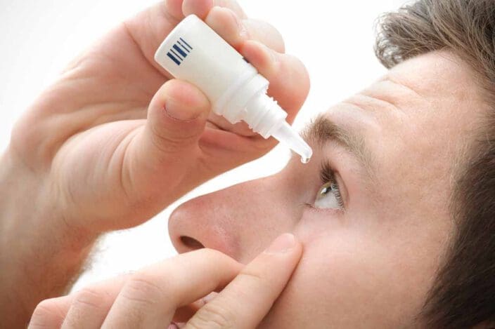
For mild hyphema, your doctor will recommend home therapies to encourage clearing the blood within a few days. Avoidance of physical activity with an elevation of the head to about 300 at night promotes reabsorption of the blood in the anterior chamber. Again, you can wear a special shield to protect your affected eye.
In some instances, the ophthalmologist will prescribe steroid eye drops to limit inflammation and swelling of the eyes. Dilating drops might also assist in reducing the discomfort of hyphema.
When receiving treatment for hyphema, you need to avoid blood thinners such as aspirin, which would promote secondary hemorrhage. If you’re taking such medications for other conditions, you should inform your doctor at the onset.
Severe cases of hyphema, grade 3 or 4, and hyphema superimposed on other eye conditions like glaucoma necessitate in-patient care for regular monitoring. Doctors prescribe eye drops containing prostaglandins, beta-blockers, alpha-adrenergic, and other components to treat hyphema with glaucoma. Oral medications also help in severe cases.
If medications do not reduce the intraocular pressures, your doctor can recommend surgical interventions such as filtering, laser therapy, drainage tube, and minimally invasive glaucoma surgery.
Hyphema Prevention
Trauma is the primary cause of hyphema. It’s therefore vital to wear protective gear for the eyes when playing contact sports such as hockey.
Prompt treatment of bleeding disorders that predispose to hyphema is also essential in preventing the condition.
You deserve clear vision. We can help.
With 135+ locations and over 2.5 million procedures performed, our board-certified eye surgeons deliver results you can trust.
Your journey to better vision starts here.
References
- What Is Hyphema? (May 19, 2021). American Academy of Ophthalmology.
- Hyphema. (September 2021). All About Vision.
- Hyphema Is Blood in the Eye. (August 11, 2021). Verywell Health.
- What Is a Slit Lamp? (April 23, 2018). American Academy of Ophthalmology.
- Glaucoma. (October 23, 2020). Mayo Clinic.
- Hyphema. (January 18, 2019). Medscape.
- Hyphema. (July 22, 2021). National Center for Biotechnology Information.
- Hyphema. (May 5, 2021). American Association for Pediatric Ophthalmology and Strabismus.
- Eye Injury Prevention. (April 14, 2021). American Academy of Ophthalmology.
This content is for informational purposes only. It may have been reviewed by a licensed physician, but is not intended to serve as a substitute for professional medical advice. Always consult your healthcare provider with any health concerns. For more, read our Privacy Policy and Editorial Policy.