Is Eye Surgery Necessary to Treat Pterygium?
Home / Surfer’s Eye (Pterygium): Signs & Removal Options /
Last Updated:
A pterygium is a type of growth on the eye. Treatment depends on the size of the growth and the symptoms that it is causing. While surgery is a treatment option, it is not always necessary.
A pterygium, also called surfer’s eye, is characterized by a bump on the eyeball that is wedge-shaped and elevated. It starts on the sclera, or white part of the eye, and over time, it can invade the cornea.
Table of Contents
This condition is not cancerous, but there is the risk for permanent issues without treatment. Visual symptoms are not uncommon.
The sun’s ultraviolet rays are the primary cause of this condition. However, other factors can contribute. Due to how this condition develops, everyone is at risk.
It is important to get an accurate diagnosis as soon as possible. The diagnostic process is straightforward and does not take long. One test is typically enough to make a definitive diagnosis.
It is important to start treatment as soon as possible after pterygium develops. This can help to reduce the risk of complications, such as scarring. In some cases, treatment is not necessary.
What Is a Pterygium?

This growth usually initiates from the eye’s corner. It occurs as a result of the body trying to protect the eye from various environmental factors, such as high levels of dust, sand, sunlight, or wind.
Anyone can develop a pterygium, but surfers are particularly prone to these growths. Because of this, it is sometimes referred to as surfer’s eye. Other at-risk groups include welders and farmers.
You deserve clear vision. We can help.
With 135+ locations and over 2.5 million procedures performed, our board-certified eye surgeons deliver results you can trust.
Your journey to better vision starts here.
While not considered to be dangerous or cancerous, some research shows that people who develop a pterygium are at a 25 percent higher risk for developing malignant melanoma. This is a type of skin cancer that can be life-threatening. Because of this, it is important to see an eye doctor any time a pterygium develops.
If a pterygium is not causing any vision problems, doctors might not recommend any treatment. However, there are treatment options when vision interference does occur.
Pterygium Symptoms
A visible growth in the corner of the eye is the most prominent symptom. This growth is painless.
People can often see blood vessels on the cornea’s outer or inner edge. In some cases, there are no symptoms associated with this condition.
However, if the pterygium becomes inflamed, people can experience irritation or burning. It can also feel like something is in the eye. If the pterygium grows into the cornea, it is possible for vision to be affected.

In some cases, people might develop a pinguecula before a pterygium. This is a small patch or bump that is yellowish in color. It occurs on the white of the eye. This precursor to surfer’s eye is generally comprised of calcium, protein, or fat deposits.
If a pinguecula is present, it can sometimes become a pterygium. It occurs as a result of chronic eye irritation.
Pterygium Causes
While this condition is not uncommon in surfers, they are not the only people at risk for developing a pterygium. There are other possible causes, such as:
- Extensive ultraviolet light exposure. Anyone who spends a lot of time in the sun can develop this condition.
- Irritant exposure. Exposure to sand, wind, and dust can increase the risk of pterygium.
- History of dry eye. Anyone who has dry eye is at a higher risk for developing this condition.
- Family history. Researchers think that if someone has family members with this condition they are at a higher risk.
Men tend to develop this issue more often than women. It also appears to be more prevalent in people between the ages of 20 and 40.
Living close to the equator increases the risk. This is due to the ultraviolet light that people are exposed to being stronger in this area of the world.
Diagnosing a Pterygium
The diagnostic process is straightforward. The doctor can use a slit lamp to make an accurate diagnosis. This examination lets the doctor see the eye with bright lighting and magnification. In most cases, this test is enough to make a definitive diagnosis.
If other tests are necessary, they may include the following:
- Corneal topography is a type of medical mapping method that doctors might use. It is done to measure the curvature of the cornea to look for changes.
- Visual acuity testing involves people reading an eye chart. The doctor will ask them to read specific lines to test their vision to see if the pterygium is interfering with vision.
- Photo documentation might be considered if the pterygium is not being treated but observed instead. It allows the doctor to track its growth.
You deserve clear vision. We can help.
With 135+ locations and over 2.5 million procedures performed, our board-certified eye surgeons deliver results you can trust.
Your journey to better vision starts here.
Less Invasive Treatment Options

- Observation: Not all pterygiums require treatment. If someone is not having discomfort or vision issues, the doctor might choose to just observe its growth. If it gets larger and starts to cause issues in the future, treatment may be considered.If this approach is taken, the doctor usually schedules examinations to occasionally look at the pterygium and evaluate for symptoms
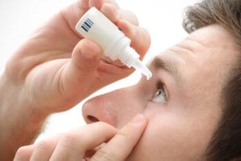
- Eye ointments or drops: If the pterygium is irritating the eye, eye ointments or eye drops might be considered. These medications alleviate redness or irritation. They reduce inflammation since they contain corticosteroids. These are used daily to decrease symptoms.
Surgical Treatments
There are surgical options if the pterygium needs to be removed. Surgery might be considered if someone has vision interference due to the growth or if eye ointments and eye drops are not alleviating the irritation.
Other reasons to consider surgery include someone not being able to move their eye normally and the appearance of the pterygium causing confidence problems.
- Auto-grafting: One procedure is called auto-grafting. This involves scraping the growth off. During this procedure, people are awake, but the doctor administers sedation to keep people comfortable during the pterygium removal.
Any conjunctiva that is removed as part of the scraping is replaced using a graft. This procedure is done the most often.
- Amniotic membrane transplantation: In some cases, the doctor might consider amniotic membrane transplantation. This is not frequently performed, but it is a surgical option. The pterygium is still scraped off like with auto-grafting. However, when the doctor replaces the conjunctiva, they use placenta instead of a conjunctiva graft.
To put the placenta piece or graft into place, the doctor uses either a fibrin glue or sutures. Sutures are considered the gold standard, but more doctors are starting to use fibrin glue.

The glue is effective, and it is simpler and faster to use. Compared to using the sutures, using glue reduces the surgical time by approximately half. It can also reduce postoperative discomfort and pain.
The recurrence rate with sutures is about 15 percent. With fibrin glue, it is about 10 to 15 percent.
- Autoblood graft fixation: This is another new approach that does not use any glue or sutures. During the procedure, the doctor excises the pterygium and the associated conjunctiva. Over the bare area, the doctor lets blood clot in a thin film. Direct tamponade is used to stop any active bleeding.
Once the clot develops, the doctor places a Tenon-free conjunctival autograft. This may or may not include limbal stem cells. Once the doctor puts the graft over the bare area, forceps are used to hold the edges in place. This is held there for about three to five minutes to allow for the fixation of the graft.
With this procedure, no recurrence of pterygium has been observed in 50 patients after four years. This is an improvement over sutures and fibrin glue.
Pterygium Prevention
Reducing exposure to the causes of this condition can decrease your risk.
Make sure to protect the eyes from wind, sunlight, and dust as much as possible. A wide-brimmed hat and sunglasses are two easy ways to accomplish this. You should also reduce your exposure to smoke and pollen whenever possible.
If someone suspects they have this condition, they should make an appointment with their eye doctor promptly. Getting an accurate diagnosis is simple and noninvasive. Then, the doctor can determine the best treatment.
You deserve clear vision. We can help.
With 135+ locations and over 2.5 million procedures performed, our board-certified eye surgeons deliver results you can trust.
Your journey to better vision starts here.
References
- Pterygia Are Indicators of an Increased Risk of Developing Cutaneous Melanomas. British Journal of Ophthalmology.
- Pterygium. MedlinePlus.
- Six Things to Know About Pinguecula and Pterygium. American Academy of Ophthalmology.
- Everything You Need to Know About Surfer’s Eye. Verywell Health.
- Slit Lamp Exam. Healthline.
- New Approach Emerges for Pterygium Surgery. EyeNet Magazine.
- Autologous Blood as Tissue Adhesive for Conjunctival Autograft in Primary Nasal Pterygium Surgery. Journal of Pre-Clinical and Clinical Research.
This content is for informational purposes only. It may have been reviewed by a licensed physician, but is not intended to serve as a substitute for professional medical advice. Always consult your healthcare provider with any health concerns. For more, read our Privacy Policy and Editorial Policy.
