
What Are the Signs of Optic Atrophy & How Do You Reverse It?
Home / Neurological Disorders /
Last Updated:
Optic atrophy is a condition that describes damage to the optic nerve leading to loss of nerve cells or tissue in the area.
Table of Contents
The optic nerve is a vital part of seeing the world around you. There are several conditions that can lead to damage to this area, including tumors, glaucoma, loss of blood flow, toxins, injury, and heredity. Types of atrophy of the optic nerve depend on where the problem starts.
If you notice any loss of vision, even if it passes, have an optometrist or ophthalmologist check your eyes. Consider getting help from a general medical practitioner too.
Any vision lost cannot be recovered, so prevention is the best form of treatment.
What Is Optic Atrophy?
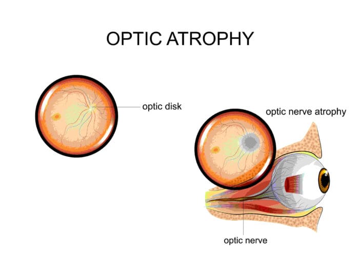
Optic atrophy, or optic nerve atrophy, is damage to the optic nerve that causes the tissues to degrade and die. This leads to a loss of vision. Light that moves through the eye hits the retina and is not processed in the brain since the signals are not transmitted by the optic nerve.
Any disease or injury that progresses to damage in the retina and the sensitive nerve behind it is considered to cause optic atrophy as a symptom, although typically a late-stage symptom.
You deserve clear vision. We can help.
With 135+ locations and over 2.5 million procedures performed, our board-certified eye surgeons deliver results you can trust.
Your journey to better vision starts here.
Atrophy of the optic nerve most often occurs in older adults, usually 60 or older. Toxins in the environment, radiation, trauma, and shock can also cause optic atrophy by harming surrounding tissues or cutting off blood flow. Blindness attributed to optic atrophy occurs in about 0.8 percent of the population.
There are some diseases of the eye and optic nerve that can cause optic atrophy too. Treatment for optic atrophy depends on the root cause.
Causes of Optic Atrophy
The optic nerve has about 1.2 million axons, originating at the ganglion cell layer, which is on the retina. These cells are myelinated, and once they are damaged in any way, they do not regenerate. Vision loss stemming from this area is typically irreversible.
There are four parts to the optic nerve:
- Intraocular part, or the optic nerve head
- Intraorbital part
- Intracanalicular part
- Intracranial part
The optic nerve is affected when optic atrophy occurs. But optic atrophy is not a disease; it is a condition that is caused by other issues.
The following issues could lead to optic atrophy:
- Brain tumor that squeezes on the area, puts pressure on the nerve or blood vessels, or infiltrates the area to cut off blood, oxygen, and nutrient supplies
- Cranial or temporal arteritis (may be called giant cell arteritis), which is inflammation of the arteries, most often in the scalp, neck, and arms, cutting off blood to these areas and leading to tissue damage
- Glaucoma, which is high pressure in the eye that leads to damage to the retina and the optic nerve
- Heredity
- Multiple sclerosis
- Stroke
Symptoms
The most common and noticeable symptom of optic atrophy is dimming vision along with reduced field of vision. This could be sudden, with the experience passing, or it could be gradual and hard to fully notice.
You may experience a dimming in your vision, which passes; you may slowly lose peripheral vision, or central vision; you may lose color vision; or you may lose spots of vision.
If you get regular eye exams, your optometrist or ophthalmologist will look at the back of your eye once a year or every two years.
Even before you lose vision, your eye doctor may notice changes to the color or size of your retina, which indicate potential damage to the optic nerve. They will then work with you to diagnose the causes of this problem and determine if you have any vision loss that you have not noticed yet.
Types of Optic Atrophy
Types of optic nerve degeneration, or atrophy, include:
- Anterograde degeneration, or Wallerian degeneration, which begins in the retina and proceeds inward. It is often caused by glaucoma.
- Retrograde degeneration, starting from the back of the optic nerve and moving toward the disc.
- Trans-synaptic degeneration, in which neurons on one side degenerate as a consequence of the loss of neurons on the other side. This is often caused by occipital damage in utero or infancy.
- Primary optic atrophy, in which optic nerve fibers die off and are replaced by glial cells without alteration in the structure of the optic nerve, causing the retinal disc to look white rather than pink-yellow.
- Secondary optic atrophy, in which optic nerve fibers degenerate with excessive proliferation and invasion of glial tissue, which dramatically changes how the area around the retina looks on eye exams.
- Consecutive optic atrophy, often due to high myopia, retinitis pigmentosa, and some other conditions, which cause the retina to turn waxy or pale and result in loss of surrounding arteries.
- Glaucomatous optic atrophy, or damage to the optic nerve caused by high eye pressure associated with glaucoma.
- Circulatory atrophy, caused by lack of blood flow to the optic nerve.
- Temporal pallor, caused by trauma to the optic nerve or malnutrition that starves the optic nerve.
Genetic heritage can also lead to optic atrophy. There are three forms of hereditary optic atrophy:
- Congenital or infantile
- Leber’s optic atrophy
- Behr hereditary optic atrophy

All three conditions start with a progressive loss of nerve cells in the retina and then progress to an atrophying of the optic nerve itself. This loss is associated with certain genetic mutations, which can be passed down in families.
Metabolic problems, including diabetes and malnutrition, can cause damage to many parts of the body, including the optic nerve.
Exposure to toxins, like tobacco, methyl alcohol, and some drugs can damage the optic nerve or cause a loss of blood flow that leads to blindness. Radiation, including from cancer treatment, can damage the optic nerve and lead to atrophy, which may be sudden or could be a slow progression over years.
You deserve clear vision. We can help.
With 135+ locations and over 2.5 million procedures performed, our board-certified eye surgeons deliver results you can trust.
Your journey to better vision starts here.
Prevention Is the Main Approach to Treating Optic Atrophy
Early diagnosis is essential to effectively managing optic atrophy and reducing the damage from it.
Preventative steps can diagnose potential optic atrophy before it starts or slow it in the early stages.
- Older adults should get regular eye exams along with blood pressure checkups.
- If your job involves toxins or physical labor, use appropriate eye protection to prevent objects or chemicals from getting into your eye.
- Get checked routinely for glaucoma, especially if you have a family history of this condition.
- Safely store chemicals in your home to prevent the spread of toxins, and never drink home-brewed alcohol as methanol can damage the optic nerve.
Tests that may be performed to determine how extensive optic atrophy is include:
- Color vision testing.
- Your pupil’s reaction to light.
- Tonometry, which measures the pressure inside your eyes.
- Visual acuity, usually through the Snellen chart.
- Visual field or peripheral vision testing.
These tests can be performed by an optometrist or ophthalmologist, but you may also need to work with your general practitioner or another medical specialist to manage underlying conditions.
Once damage to the optic nerve occurs and vision is lost, it cannot be recovered. Medical treatments to slow the progression of any underlying chronic conditions, or to stop damage to nerves and blood vessels, will slow or stop the progression toward blindness.
It is also important to protect the other eye during treatments involving optic atrophy. While many conditions do not damage the eyes symmetrically, chronic health problems can begin to damage the other eye after the first optic nerve begins to atrophy.
Living With Blindness Due to Optic Atrophy
Since there is no cure for optic atrophy, people who experience vision loss from it need support as they adjust. Adaptive strategies can help you cope.
- Get organized. A friend or family member can help you organize your home so essential items are easily found and within reach.
- Consider contrast. Some people are still able to differentiate between dark and light colors if they experienced vision loss rather than blindness. Use contrasting colors to help identify certain items.
- Create paths. Walkways throughout the home should be clear and free of clutter to avoid trips and other hazards.
- Build a support system. Vision loss, including blindness, is a huge life change that comes with physical and emotional challenges. Reach out for support from loved ones and support groups. The American Council for the Blind offers resources for people living with blindness and vision loss.
You deserve clear vision. We can help.
With 135+ locations and over 2.5 million procedures performed, our board-certified eye surgeons deliver results you can trust.
Your journey to better vision starts here.
References
- Optic Nerve Atrophy. (April 2019). MedlinePlus.
- Optic Atrophy. (August 2021). StatPearls.
- Optic Atrophy. (February 2019). EyeWiki, American Academy of Ophthalmology (AAO).
- Giant Cell Arteritis. (January 2019). MedlinePlus.
- Hereditary Optic Nerve Disorders. (July 2018). Merck Manual, Consumer Version.
- Tonometry. (April 2019). MedlinePlus.
This content is for informational purposes only. It may have been reviewed by a licensed physician, but is not intended to serve as a substitute for professional medical advice. Always consult your healthcare provider with any health concerns. For more, read our Privacy Policy and Editorial Policy.
